Description
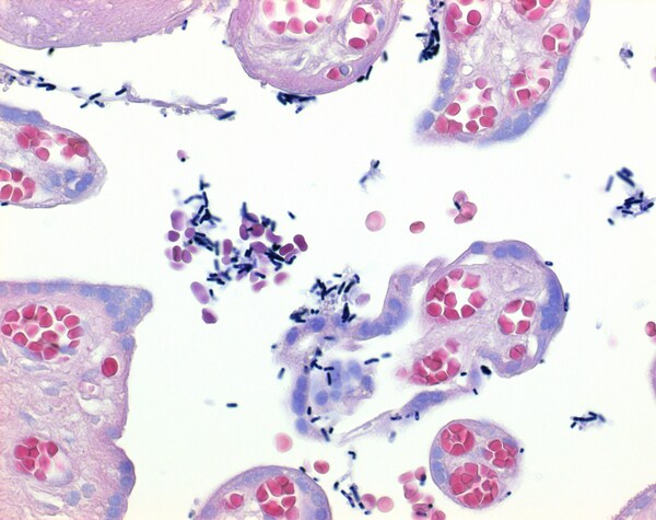
The Gram staining allows a fast differentiation of bacteria in Gram-positive and Gram-negative. The mureine structure of the bacteria walls is the basis of the color affinity. Bacteria will be stained with Gram’s crystal violet solution – an aniline dye – in the first step. After the treatment with iod solution (Lugol’s solution), a dye-iod complex will form. During the decolorizing step, this complex stays in the multilayer mureine structures of the Gram-positive bacteria and they will appear blue. Gram-negative bacteria have a monolayer mureine structure only, the dye-iod complex does not stay bound to the cellwall, they will be decolorized . Gram-negative bacteria will be counterstained by safranin solution and appear orange. A bottle with 500 ml Gram’s crystal violet solution is sufficient for staining up to 250 slides.


Reviews
There are no reviews yet.